Sleep, Biological Rhythms & Cellular Metabolism
BIOMARKERS
IL-1 Beta
TNF-Alpha
IL-10
REM – Rapid Eye Movement
Melatonin
BDNF – Brain Derived Neurotrophic Factor
Sleep-Disordered Breathing
Seratonin
Noradrenalin
Dopamine
Acetylcholine
PAT – Peripheral Arterial Tone
EQUIPMENT
Features: AS40 amplifier with 35 channels of data, sampling rate of 800 samples/second/channel. Data stored at 200 or 400 samples/second, user definable via TWin software. The lab-based recorder includes PC, keyboard and mouse, DVD+/RW Archiver (internal), 10/100 Base-T NIC and modem. Also includes TWin PSG Record and Review, Archive Managing software, TWin Companion Holter EKG software, MS Office software. 35-channel AS40 Amplifier System with tethered Sleep Personality Module, Personality Module Adaptor, and Power Supply. DCM8 DC Input Module for up to 8 channels, and 20-button programmable keypad.This state-of-the-art monitoring system is powerful and easy to use, providing data acquisition, recording and review capabilities in one flexible package. The Comet systems provide for patient safety isolation, signal conditioning and digitization.[/column]
 The Grass SPM sleep diagnostic headbox provides 30 AC channels and can be worn by the patient or conveniently placed bedside. The compact headbox is especially useful with the Comet PSG system which includes all the latest features for AASM requirements. The powerful bedside amplifier and fully featured TWin software make Comet the leading choice of sleep professionals.
Stage and score recordings efficiently and accurately. A predefined list of stages and scores can be utilized, or create new events to meet your specific needs. Computer assisted sleep scoring is available for Respiratory, Leg Movement, Snore, Bruxism, and Desaturation events. Automatic Arousal correlations and EKG, body position, EtCO2, TCO2, and DC channel trending is also available. Finally, utilize the Sleep Scoring Comparison Module to assist with laboratory inter-scorer reliability comparisons.[
Neurotrac-III Neuromonitoring Software is designed for computing and displaying long-term trends of EEG features during continuous EEG monitoring in the ICU, NICU, OR or Seizure Monitoring units. The software module can be used with acquisition systems configured with any of Grass Technologies’ amplifiers. The number of EEG channels recorded is dependent on the amplifiers used, the electrodes applied to the patient, and the selected montage. Systems configured with the Neuromonitoring software can also be used for routine EEG, LTM and PSG by adding Photic Stimulators, Digital Video options, SzAC online seizure/spike detection, Auto-Sleep Analysis and FASS
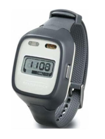
WatchPAT is an FDA-approved diagnostic device that uses innovative technology to ensure the accurate detection of sleep apnea. Its ease of use is unparalleled and it is greatly complemented by the fact that WatchPAT testing is done in the comfort of your own bedroom; an environment that best reflects the pattern of your sleep habits. WatchPAT monitors changes in peripheral arterial tone and activity, as well as in blood oxygen saturation levels. It also identifies sleep apnea events just like the equipment used in polysomnography (PSG) sleep studies performed in hospital sleep laboratories.
 In addition to sleep/wake activity recording and the updated features included in the Actiwatch-2, Actiwatch Spectrum is equipped with three color light sensors that provide irradiance and luminous flux recordings in three color bands of the visible spectrum: red, green, and blue. Beyond color-light sensing, Actiwatch Spectrum also features a LCD display for time, date and device status indicators as well as off-wrist detection and a one year battery life. These features, among others are why Respironics’ Actiwatch products make it easier than ever to implement actigraphy.
CLINICAL SIGNIFICANCE
Polysomnography (PSG) is a comprehensive recording of the biophysiological changes that occur during sleep. PSG monitors many body functions including brain (EEG), eye movements (EOG), muscle activity (EMG), and heart rhythm (ECG), respiratory airflow and pulse oximetry. PSG is used to diagnose sleep disorders including narcolepsy, periodic limb movement (PLMD), REM disorder and sleep apnea.
IL-1Beta and TNF-Alpha are involved with NREM regulation and some of the symptoms of sleep loss. ATP plays a part in the release of IL-1 and TNF.
Melatonin (an anti-oxidant secreted during sleep) is produced by the pineal gland located in the center of the brain outside of the blood-brain barrier. The Melatonin signal forms part of the system that regulates the sleep-wake cycle. During sleep the release of certain neurotransmitters, the monamines (norepinephrine, serotonin and histamine) is completely shutdown. The functions of REM (rapid eye movement) sleep include memory, brain development, and dreams. During the stages of NREM sleep, the body repairs and regenerates tissue, builds bone and muscle and appears to strengthen the immune system.
BIOMARKERS
Melatonin
Core Body Temperature (CBT)
Blood Plasma Cortisol Levels
Human Growth Hormone (HGH) Levels
Heart Rate
EQUIPMENT
 The Actical’s small size, waterproof case, and ability to be worn in multiple locations makes it easy for subjects to wear regardless of their activity. The device is positioned securely on the subject using a wristband, waistband, or ankle band. Once the device is on, subjects can wear it and forget it and go about their normal daily activities, including rigorous exercise, swimming or bathing. There is no need to worry about subjects repositioning, damaging, or removing the device. Now with 32MB of on board memory, and high speed sampling capabilities, the Actical system makes monitoring physical activity for days or weeks a practical reality. Powerful, fast, and flexible, Actical helps you collect valuable physical activity and energy expenditure data simply and reliably. The Actical physical activity monitoring system is a rigorous solution designed with the needs of serious researchers in mind.
Activity is a standard marker of circadian rhythms in studies of non-human mammals. This section examines the use of wrist activity in the measurement of circadian rhythms in humans. In the studies reviewed here, wrist actigraphy was used in a number of different ways relevant to human rhythms. Methodologies included characterizing spontaneous rhythms in adults, children, infants and the elderly, exploring the relationships between activity rhythms and the light-dark cycle, helping to identify sleep or rhythm disturbances induced by change of schedule, measuring improvement in disturbed rhythms after experimental intervention, helping to diagnose circadian rhythm sleep disorders, characterizing rhythm abnormalities that accompany dementia or psychiatric disturbance, and investigating the role of motor activity in cardiovascular rhythms.
 Core body temperatures are obtained from ingestible and, of course, disposable, Jonah core temperature sensors, which are contained in a capsule about the size of a multi-vitamin. It weighs only 1.6 grams and measures 8.7 mm dia. x 23mm long and is made of medical grade plastic. Once activated and swallowed, transmission begins immediately. Data are transmitted telemetrically to the VitalSense Monitor, which can be worn in a waist pack or slipped into a pocket. The mean transit time for the capsules is 2.0 ±1.5 days. Dermal temperatures are transmitted from waterproof, hypoallergenic patches. Transmission range is approximately 1 meter for the ingestible sensor and 2 meters for the patches. The patented redundant transmission scheme for the sensors significantly decreases the number of lost data points. Accuracy is ±0.1 °C with 0.01 ºC resolution.
CLINICAL SIGNIFICANCE
Elevated melatonin levels are correlated with the sleep-wake cycle but also play an important role in the recognition of the light-dark cycle in organisms. Melatonin levels can be measured in the blood, saliva, or through urine upon the awakening of the subject.
Core Body Temperature (CBT) typically fluctuates between 37-37.6 °C during daytime hours of work or activity. Upon entering a state of sleep or rest in the evening, between midnight and 8am, the CBT drops to a range of 32.2-32.6 °C due to the decreased metabolic rate and lack of need for energy. VitalSense proved to be a real lifesaver in a recent study of wildlandfirefighters in Montana. The study was designed to evaluate heat stress in high intensity work environments. Canadian coaches used VitalSense to evaluate the physiological status of Canadian triathletes training for all three legs (swimming, biking and running) of their event in the 2004 summer Olympics.
In another athletic related study, Nike is using VitalSense to test heat dissipation in clothing.
VitalSense has been incorporated into a number of on-going clinical studies. Those in early stages of design and testing include menopausal hot flash monitoring, ovulation detection, and sepsis detection in hospitals.
Hormonal levels of cortisol and human growth hormone (HGH) also experience cyclic fluctuations. During the sleep or resting state, cortisol levels decline significantly and HGH levels increase; this is due to the shift in priority from energy mobilization to energy conservation and tissue growth/reparations.
Heart Rate also declines synchronically with CBT and hormonal levels due to the reduced need for energy and aerobic respiration.
BIOMARKERS
Temperature
Core Temperature
Peripheral Temperature
Inflammation
C-Reactive Protein (CRP)
Pro-Inflammatory
Hydrogen Peroxide (H2O2)
Histamine
Tumor Necrosis Factor Alpha (TNF-a)
Interleukin 6 (IL-6)
Interleukin 1 Beta (IL-1b)
Interleukin 12 (IL-12)
Interferon Gamma
Anti-Inflammatory
Interleukin 4 (IL-4)
Interleukin 10 (IL-10)
Interleukin 16 (IL-16)
Interferon Alpha
EQUIPMENT
 Core body temperatures are obtained from ingestible and, of course, disposable, Jonah core temperature sensors, which are contained in a capsule about the size of a multi-vitamin. It weighs only 1.6 grams and measures 8.7 mm dia. x 23mm long and is made of medical grade plastic. Once activated and swallowed, transmission begins immediately. Data are transmitted telemetrically to the VitalSense Monitor, which can be worn in a waist pack or slipped into a pocket. The mean transit time for the capsules is 2.0 ±1.5 days. Dermal temperatures are transmitted from waterproof, hypoallergenic patches. Transmission range is approximately 1 meter for the ingestible sensor and 2 meters for the patches. The patented redundant transmission scheme for the sensors significantly decreases the number of lost data points. Accuracy is ±0.1 °C with 0.01 ºC resolution.
CLINICAL SIGNIFICANCE
 TEMPERATURE
VitalSense proved to be a real lifesaver in a recent study of wildland firefighters in Montana. The study was designed to evaluate heat stress in high intensity work environments.
Canadian coaches used VitalSense to evaluate the physiological status of Canadian triathletes training for all three legs (swimming, biking and running) of their event in the 2004 summer Olympics.
In another athletic related study, Nike is using VitalSense to test heat dissipation in clothing.
VitalSense has been incorporated into a number of on-going clinical studies. Those in early stages of design and testing include menopausal hot flash monitoring, ovulation detection, and sepsis detection in hospitals.
INFLAMMATION
Inflammation is the body’s response to injury or infection it can be classified as either acute or chronic. Acute inflammation is the initial inflammatory response it occurs almost immediately after minor injuries like burns and cuts as well as major trauma such as myocardial infarction (MI). Acute inflammation results in the healing of the tissue when the injury or infection is removed. Symptoms include redness, swelling, heat, pain and stiffness in the affected area. Chronic inflammation or prolonged inflammation may follow acute inflammation or exist independently. Chronic inflammation is a continuous process for example tissue breakdown and repair attempts that often results in scarring and tissue destruction. Chronic inflammation can arise after bacterial infection or as a result of an autoimmune disease. In autoimmune diseases, the inflammatory response is triggered when there are no stimuli and the immune system attacks itself.
Extra protein is often released from the site of inflammation these proteins can be readily detected in the bloodstream and are therefore referred to as inflammatory markers. Perhaps the most commonly used marker of inflammation is C-reactive protein (CRP).
BIOMARKERS
Regional Oxygen Saturation (rSO2)
Oxyhemoglobin
Deoxyhemoglobin
Total Hemoglobin
Oxygen Content
Carboxyhemoglobin
Methemoglobin
EQUIPMENT
 The INVOS system is the only cerebral/somatic oximetry system with FDA cleared improved outcome claims. The INVOS system provides real-time monitoring of changes in regional oxygen saturation (rSO 2) of blood in the brain or other body tissues beneath the sensor for effective oxygen monitoring in adults, children, infants and neonates. This unique system allows clinicians to measure site-specific oxygen levels rather than requiring them to infer the data from systemic, whole body measures such as blood pressure and pulse oximetry.
Available in two or four data channels, clinicians can conveniently monitor multiple brain and body areas. The INVOS system uses near-infrared light at wave-lengths that are absorbed by hemoglobin (730 and 810 nm). Light travels from the sensor’s light emitting diode to either a proximal or distal detector, enabling separate data processing of shallow and deep optical signals. The INVOS system’s ability to localize the area of measurement, called spatial resolution, has been empirically validated in human subjects. Data from the scalp and surface tissue are subtracted and suppressed, reflecting rSO2 in deeper tissues.
 Featuring “gold standard” Masimo SET pulse oximetry and proven in more than 100 independent and objective studies to provide the most accurate and reliable SpO2 readings during motion and low perfusion. An upgraded Masimo Rainbow SET technology platform allows continuous and noninvasive measurement of oxygen saturation, pulse rate, and perfusion index in addition to total hemoglobin, total arterial oxygen content, PVI, carboxyhemoglobin, and methemoglobin.
A pulse oximeter works by passing a beam of red and infrared light through a pulsating capillary bed. The ratio of red to infrared blood light transmitted gives a measure of the oxygen saturation of the blood. The oximeter works on the principle that the oxygenated blood is a brighter color of red than the deoxygenated blood, which is more blue-purple. First, the oximeter measures the sum of the intensity of both shades of red, representing the fractions of the blood with and without oxygen. The oximeter detects the pulse, and then subtracts the intensity of color detected when the pulse is absent. The remaining intensity of color represents only the oxygenated red blood. This is displayed on the electronic screen as a percentage of oxygen saturation in the blood.
CLINICAL SIGNIFICANCE
Cerebral oximetry is the measurement of oxygen saturation in the brain. This organ requires a great deal of oxygen to function and is extremely sensitive to periods of deprivation. Monitoring oxygen levels can provide important information about a patient’s neurological health, and may allow care providers to rapidly address falling oxygen saturation in the brain. This can improve patient outcomes and reduce the risk of brain damage, stroke, and other neurological trauma. In addition to being used in surgery, cerebral oximetry can have some other applications. Sleep studies may involve the use of oximetry to evaluate oxygen levels in the brain and elsewhere in the body to determine if patients experience oxygen deprivation during sleep.
Oximetry is a procedure for measuring the concentration of oxygen in the blood. The test is used in the evaluation of various medical conditions that affect the function of the heart and lungs. Pulse oximeters are used by anesthesiologists as the universal standard of care in operating theatres, emergency, recovery and neonatal units and all wards (especially in pediatric wards). Oximeters are used in the treatment of pneumonia, used to prevent neonatal blindness and are a key component of the WHO Safe Surgery Checklist.


